Early Detection of Breast Cancer
Here are some ways your health care provider can find breast changes of the body.

Clinical breast exam
During a clinical breast exam, your health care provider checks your breasts and nipples and under your arms for any abnormal changes.
Ask your health care provider at what age and how often you should have a clinical breast exam. During the visit, it’s important to share your personal medical history and your family medical history. This includes problems or diseases that you or your family members have had.
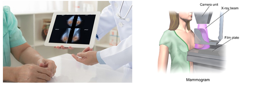
Mammogram
A mammogram is an x-ray picture of your breast tissue. This test may find tumors that are too small to feel. During a mammogram, each breast is pressed between two plastic plates. Some discomfort is normal, but if it’s painful, tell the mammography technician.
Film mammography stores the image directly on x-ray film.
Digital mammography takes an electronic image of the breast and stores it directly in a computer. Digital images can be made lighter, darker, or larger. Images can also be stored and shared electronically.
Understanding Your Mammogram Report
A doctor called a radiologist will categorize your mammogram results using a numbered system. Talk to your doctor about your mammogram results and what you need to do next.
BI-RADS
Breast Imaging Reporting and Data System or BI-RADS is the standard method for doctors to describe mammogram findings and results. It consists of six categories that allow doctors to identify what the results of a mammogram mean.
“BI-RADS 1 means that the mammogram was negative (i.e., no cancer) and that you should continue your routine screening. BI-RADS 2 also means that your mammogram was normal (i.e., no cancer), but other findings (e.g., cysts) are described in the report.”
Read More – https://www.breastcancer.org/screening-testing/mammograms/bi-rads-results
https://www.nationalbreastcancer.org/early-detection-of-breast-cancer/
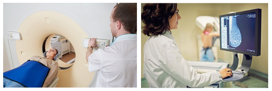
Magnetic resonance imaging, also called MRI, uses a powerful magnet, radio waves, and a computer to take detailed pictures of areas inside the breast. MRI is another tool that can be used to find breast cancer. However, MRIs don’t replace mammograms. They are used in addition to mammograms in women who are at increased risk of breast cancer.
MRIs have some limits. For example, they cannot find breast changes such as microcalcifications. MRIs are also less specific than other tests. This means that they may give false-positive test results—the test shows that there is cancer when there really is not.
Breast MRI may be useful in some special situations.
- Breast cancer screening for young women who have an increased risk of breast cancer, especially in those with strong family history or in those with mutations in the genes BRCA1 or BRCA2.
- Evaluation for breast cancer in a woman who is diagnosed with cancer of the lymph nodes (glands) under the arm but who has no sign of breast cancer in either breast. Breast MRI can be used to determine the exact location of the cancer in the breast.
- Evaluation of a woman with newly diagnosed breast cancer with extremely dense breasts on mammograms, because the density of the breast tissue makes the mammograms difficult to interpret.
What Our Patients Are Saying About Us
